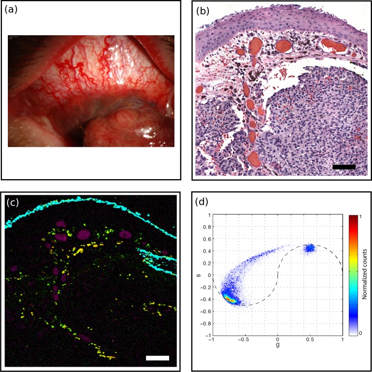
Figure 4.
Melanoma (MM1). (a) A 61-year-old male presented with pigmented conjunctival lesion enlarging over several years. Marked vascularity of the lesion is evident clinically. (b) H&E-stained histology section, scale bar: 100 μm. Intraepithelial melanocytic hyperplasia is noted on histology while nests of atypical melanocytes invade substantia propria. Marked vascular congestion is also noted. (c) Pump-probe image demonstrated distribution of eumelanin (red) and pheomelanin (green) pigment, predominantly located near vasculature (magenta). Surgical ink in cyan. Scale bar: 100 μm. (d) Associated phasor plot.

