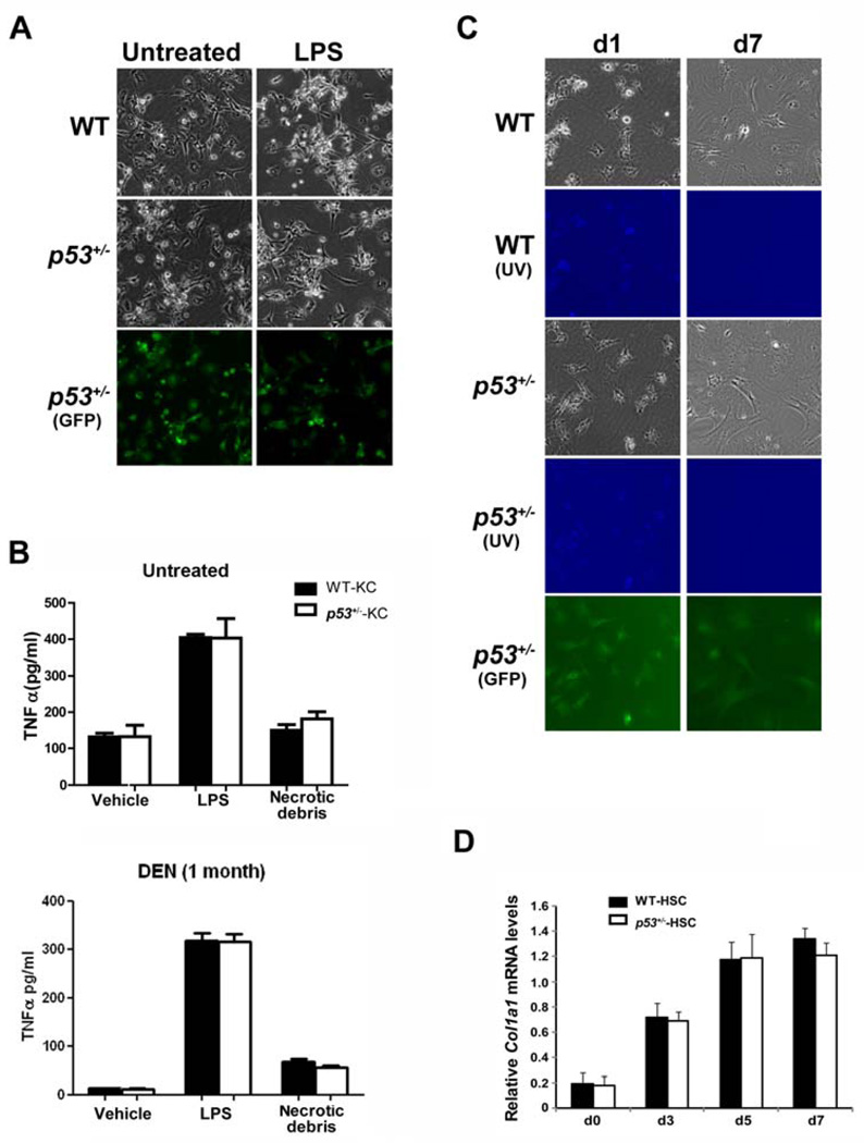
Figure 1. p53 deficiency does not affect the innate inflammatory response of non-parenchymal cells in liver.
(A–B) Kupffer cells were isolated from untreated or DEN-treated (i.p. injection with 70 mg/kg DEN weekly for one month) WT and p53+/− rats of the same littermate and treated with lipopolysaccharide (LPS, 10 ng/ml) or necrotic cellular debris prepared by cycles of freeze-thawing of primary hepatocytes for 16 h. Following treatment, cultures were analyzed for morphological evidence of Kupffer cell activation (A) and TNFα production in the media using ELISA (B).
(C–D) Quiescent hepatic stellate cells from WT and p53+/− rats were cultured on plastic for 7 days. Activation of stellate cells is characterized by the development of fibroblastic morphology as well as loss of intracellular vitamin A content as assessed by UV-excited autofluorescence (UV) (C), concomitant with induction of the HSC activation marker Col1a1 (D). Data are presented as mean ± s.d. Cells isolated from heterozygous p53tm1(EGFP-pac) rats (p53+/− rats) express GFP as a conceptual proof.

