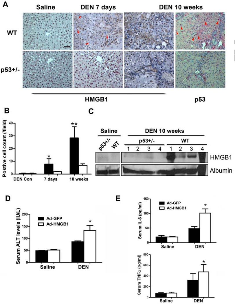
Figure 3. Secreted HMGB1 contributes to DEN-induced chronic hepatic injury and tumorigenesis.
(A) Immunohistochemistry analysis of HMGB1 and p53 in liver sections of WT rats compared with p53+/− rats 7 days or 10 weeks after DEN treatment. Red arrowheads indicate areas showing cytoplasmic translocation of HMGB1 or nuclear accumulation of p53. Scale bars, 50 µm.
(B) Quantification of cells positive for cytoplasmic HMGB1 in liver sections of (A). Results are mean±s.d., *P<0.05, **P<0.01.
(C) Circulating levels of HMGB1 were measured by immunoblotting in WT and p53+/− rats on week 10 of DEN treatment. Albumin in serum was blotted as loading control. Numbers indicate individual rats.
(D) Wild-type rats were i.v. injected with recombinant adenoviruses expressing GFP or HMGB1 and, 24h later, were treated with saline or DEN. Forty eight hours later, serum levels of ALT were measured. Results are means ± s.d., n = 5 in each group. p = 0.0345 for ALT
(E) Serum levels of IL-6 and TNFα were measured in rats treated in (D). Results are means ± s.d., n = 5 in each group. and p= 0.0163 for IL-6 levels and p = 0.0333 for TNFα levels, respectively.

