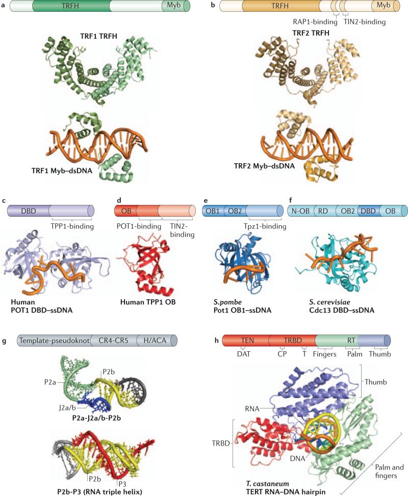
Figure 2.
Structures of telomeric proteins and telomerase components. a, Crystal structure of TRFH domain (PDB: 1H6O) and dsDNA-bound Myb domain of human TRF1 (PDB: 1W0T). b, Crystal structure of TRFH domain (1H6P) and dsDNA-bound Myb domain of human TRF2 (PDB: 1W0U). c, Crystal structure of ssDNA-bound DNA-binding domain (DBD) of human POT1, comprised of two OB folds (PDB: 1XJV). d, Crystal structure of the N-terminal OB domain of human TPP1 (PDB: 2I46). e, NMR structure of ssDNA-bound DBD of S. cerevisiae Cdc13 (PDB: 1S40). f, Crystal structure of ssDNA-bound structure of the first OB domain (OB1) of S. pomble Pot1 (PDB: 1QZG). g, NMR structures of template-pseudoknot (PK) fragments of human TR containing the indicated secondary structural elements; top structure (PDB: 2L3E); bottom structure (PDB: 2K96). The ‘P’ and ‘J’ elements are RNA double-helical paired regions and joining segments, respectively. h, Crystal structure of T. castaneum TERT with a hybrid RNADNA hairpin representing a putative telomerase-primer-template ternary complex (PDB: 3KYL).

