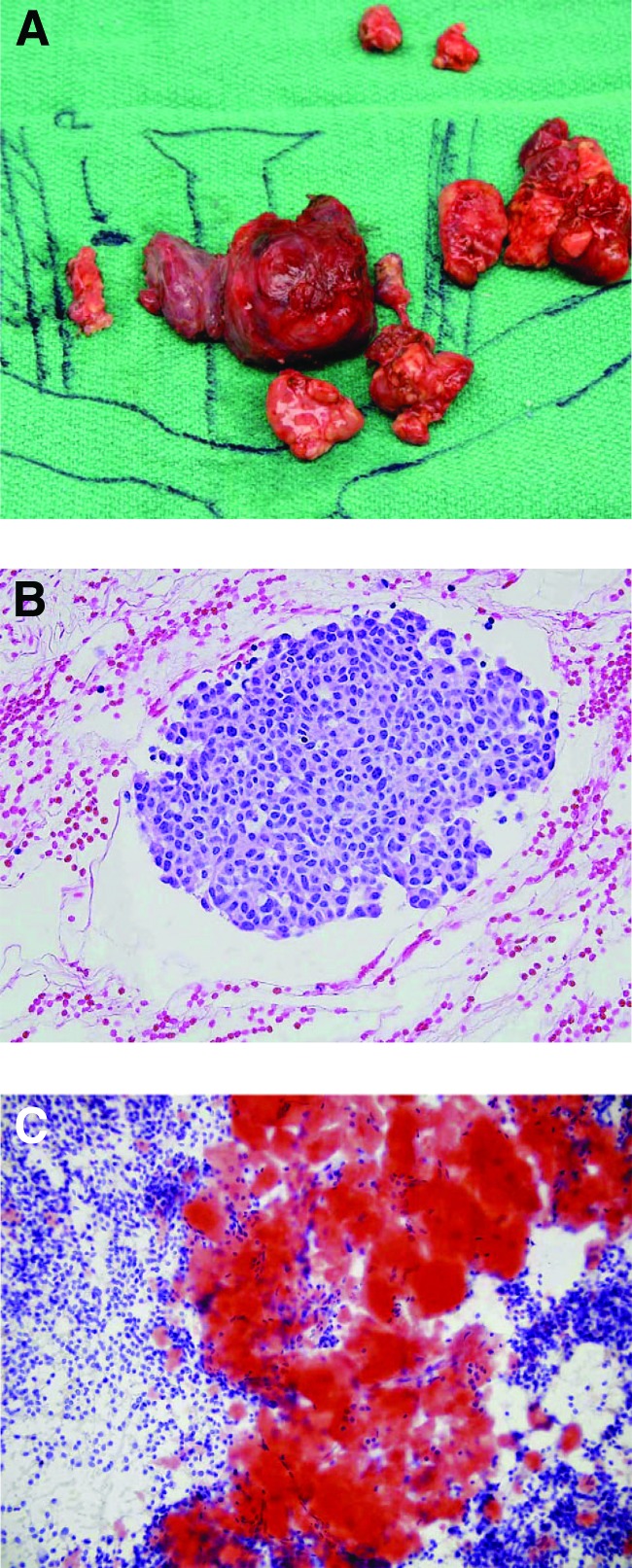
Figure 1.
Gross view and histology of medullary thyroid cancer. (A): Operative specimen of left thyroid and associated lymph nodes. (B, C): Histology of medullary thyroid cancer shown by hematoxylin and eosin (B) and Congo red stain (C) emphasizing the characteristic stromal amyloid. From [41] with permission from AlphaMed Press.

