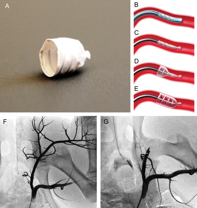
Figure 1:
EOS device, illustration of the EOS occlusion mechanism and angiography before and after EOS device deployment. EOS device shown in deployed configuration (A). The EOS is delivered through the guide sheath into the target position of the vessel (B) before the EOS device is unsheathed for deployment (C). The deployment process is initiated by release of the proximal end (Step 1). At this stage, the operator is still in full control to refine the exact target position (D). Finally, the distal end of the implant is deployed (Step 2), leading to immediate vessel occlusion. Thereafter, the carrier catheter can be easily removed (E). Predeployment angiography was performed to define the target vessel and to assess the vessel diameter to determine the optimal EOS device size (F). After deployment of the EOS device, immediate and complete vessel occlusion was achieved (G)

