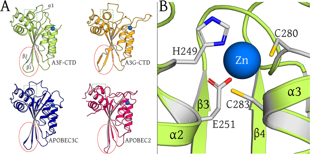
Fig. 3. Structural Alignment of APOBEC family members.
(A): Comparison of ribbon representations of A3F-CTD, A3G-CTD (3V4K), A3C (3VOW) and APOBEC2 (2NYT) crystal structures (chain A of all). The β2/ β2-β2’ region is circled in red to highlight similarities across all proteins except A3G-CTD. The α1 helix region is boxed in green to highlight similarities across all proteins except APOBEC2.
(B): The catalytic zinc (blue sphere) is coordinated at the active site by H249, C280, C283 and indirectly via a water molecule (view occluded by zinc atom) with E251.

