
Figure 1.
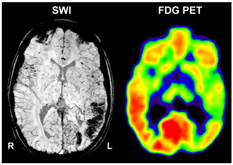
Co-registered susceptibility weighted imaging (SWI) and 2-deoxy-2[18F]fluoro-D-glucose positron emission tomography (FDG-PET) in a child with Sturge–Weber syndrome and left hemispheric involvement. Native SWI shows calcified areas in the left occipital and posterior temporal cortex. Hypometabolism on PET extends into the anterior temporal cortex.