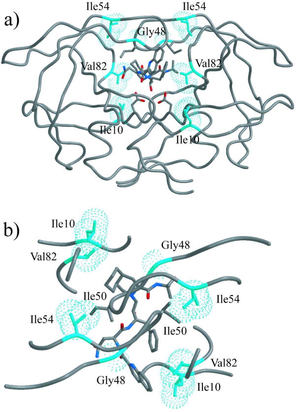
Figure 3.

Positions of mutated residues displayed on inhibitor-bound HIV-1 protease structure (WT protease bound to SQV, PDB code 1HXB). Ile10, Gly48, Ile54, and Val82 side chains are in cyan van der Waal spheres. The SQV molecule is in stick representation and colored according to atom type, with grey for carbon, blue for nitrogen, and red for oxygen. (a) Front view, showing that neither residue 10 nor residue 54 is in contact with the inhibitor. (b) Top view, showing that residue 54 is in close contact with Ile50 of the other monomer.
