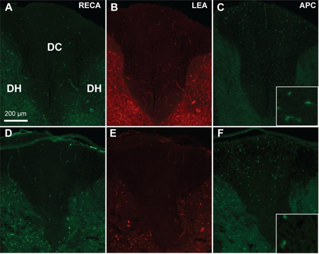
Figure 3. Contusive SCI leads to acute microvascular and oligodendrocyte loss.
A 120 kdyn contusion at T9 leads to loss of total (RECA, D) and perfused (LEA, E) microvessels in the dorsal column compared to sham (A,B). Shown here are sections at 1mm rostral to the epicenter from representative rats at 6 hr post-injury. Similarly, APC+ oligodendrocyte loss is observed after contusion (F) compared to sham (C) in adjacent sections. Insets are 63× objective images APC+ cell bodies. Scale bar 200 µm.

