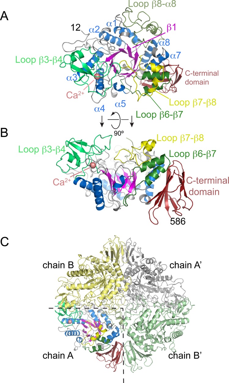
Figure 2.

Overall fold of M. tuberculosis trehalose synthase TreS and its tetrameric assembly in the crystal. (A) Top view of the structure, with a (β/α)8-barrel fold (blue helices, magenta strands) as the conserved core and an antiparallel β-sandwich domain at the C-terminus (dark red). Selected loops connecting successive β-strands in the (β/α)8-fold are highlighted. (B) Orthogonal view of panel A. (C) Quaternary structure of TreS, containing 2 copies of each of chain A and B. Primes denote copies linked by crystallographic symmetry.
