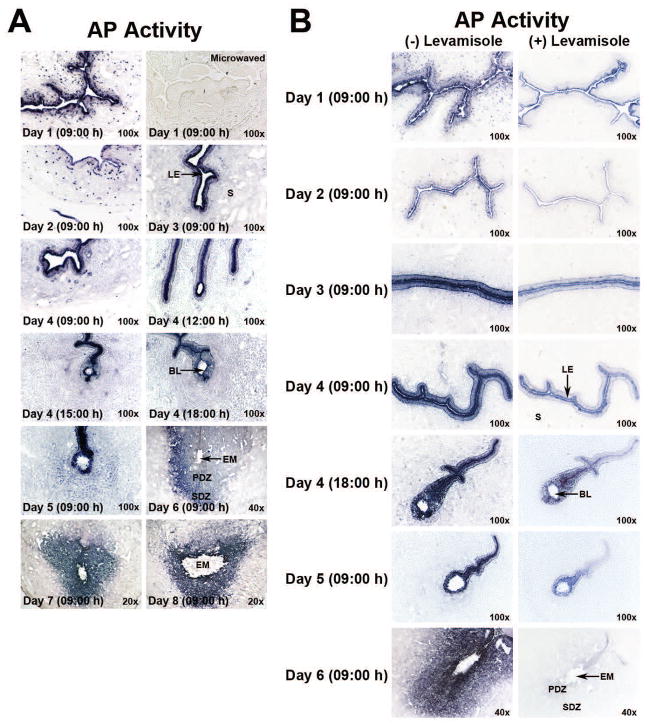
Fig. 5.
Total and levamisole-sensitive and –insensitive AP activities in the periimplantation hamster uterus. Cross-sections from days 1 to 4 uteri and days 4 to 8 implantation sites were processed to demonstrate cell-specific localization of total AP (A) and levamisole-insensitive AP (B) activities. Photographs were captured under bright-field (n=3 or 4/day of pregnancy). BL, Blastocyst; EM, Embryo; LE, Luminal Epithelium; PDZ, Primary Decidual Zone; S, Stroma; SDZ, Secondary Decidual Zone. A) Total AP activity in periimplantation uteri (days 1–8) as determined by histochemistry. Specificity of AP staining in sections from day 1 uteri was demonstrated by destroying endogenous enzyme activity through microwave heating. B) Residual AP (levamisole-insensitive) activity after levamisole treatment in the periimplantation (days 1–6) uterine sections.

