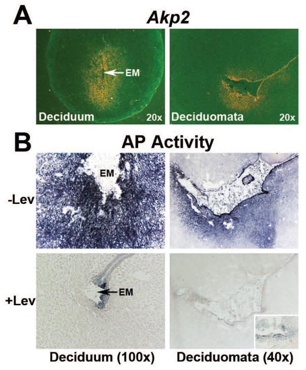
Fig. 6.
Expression of Akp2 mRNA (A) and total AP activity (B) in the day 6 embryo-induced deciduum and suture-induced deciduomata. BL, Blastocyst; EM, Embryo. A) Expression of Akp2 mRNA by in situ hybridization. Photographs were captured under dark-field (n=3). B) Histochemical staining of AP activity in the presence and absence of levamisole (Lev). Photographs were captured under bright-field (n=4). Inserts shows higher magnification (200x) of levamisole-insensitive AP activity in residual epithelial cells.

