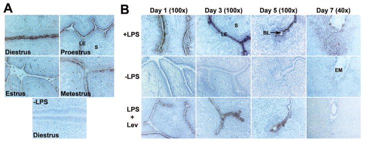
Fig. 8.

LPS dephosphorylation at uterine sites of AP production during the cycle and periimplantation period. Histochemical localization of AP activity in sections from cyclic (A) and periimplantation uteri (day 1, day 3 and implantation sites of days 5 and 7) (B) using LPS as a substrate. All sections were counterstained with hematoxylin. A dark brown staining indicated lead sulphide precipitates. Photographs were captured under bright-field (n=4). Control sections without LPS were completely negative. Residual stainings in levamisole (Lev)-treated sections from day 1, day 3 and day 5 uteri indicated levamisole (Lev)-insensitive gIAP activity. BL, Blastocyst; EM, Embryo, LE, Luminal Epithelium; S, Stroma.
