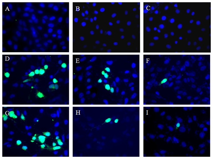
Figure 2. Immunofluorescence studies of HSV-2 infected cells treated with HM.
Cells treated with HM at 2-4 h post-infection (p.i) were fixed with paraformaldehyde and blocked with BSA-PBS-triton X100 solution. After permeabilization, cells were incubated with FITC-labelled polyclonal rabbit anti-HSV-2 antibody, and observed under Axio Imager M1 (Carl Zeiss, NY, USA) inverted epifluorescence microscope. Cell Control [a]; Cells treated with HM (5.0 μg/ml) at 2 h [b] and 4 h [c] p.i.; Virus control at 2 h p.i [d]; infected cells treated with HM at 1.5 μg/ml [e], and 5.0 μg/ml [f] at 2 h p.i; Virus Control at 4 h p.i [g]; infected cells treated with HM at 1.5 μg/ml [h] and 5.0 μg/ml [i] at 4 h p.i.

