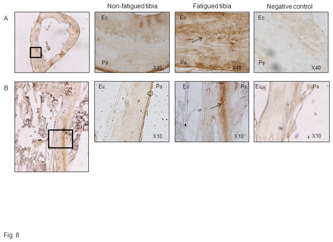
Figure 8. Fatigue loading stimulates periostin expression in cortical bone.
Immunohistochemical staining of periostin expression in cross sections (A) and longitudinal sections (B) of the loaded and non-loaded midshaft tibia 3days after fatigue. Magnified images of the cortical region underlying soft calluses are also shown. Note the presence of periostin into osteocyte canaliculae of the fatigued tibia (A, arrow), as well as in the periosteum (Ps) of fatigued tibiae, whereas weak or no staining is detectable at endocortical surfaces (Ec).

