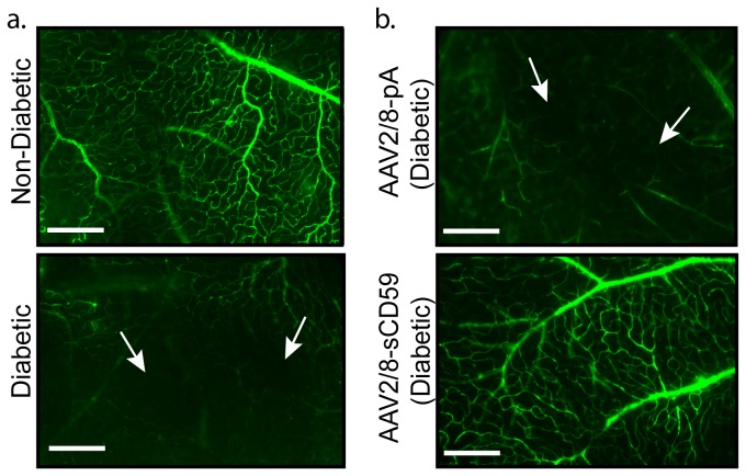
Figure 2. sCD59 Attenuates Non-Perfusion of the Retinal Vessels in Diabetic Mice.
(a). Representative images of retinal flat-mounts from un-injected diabetic eyes (n=7) and un-injected non-diabetic eyes (n=4), following intra-cardiac perfusion with fluorescein-dextran. Retina from un-injected diabetic mouse shows areas of capillary non-perfusion (white arrows). Scale bars = 120μm. (b). Representative images of retinal flat-mounts following intra-cardiac perfusion of fluorescein-dextran shows attenuation of areas of non-perfusion of retinal vessels in AAV2/8-sCD59 injected diabetic eyes (n=6), while AAV2/8-pA injected diabetic eyes (n=4) show areas of capillary non-perfusion (white arrows). Scale bars = 120μm. n represents the number of eyes.

