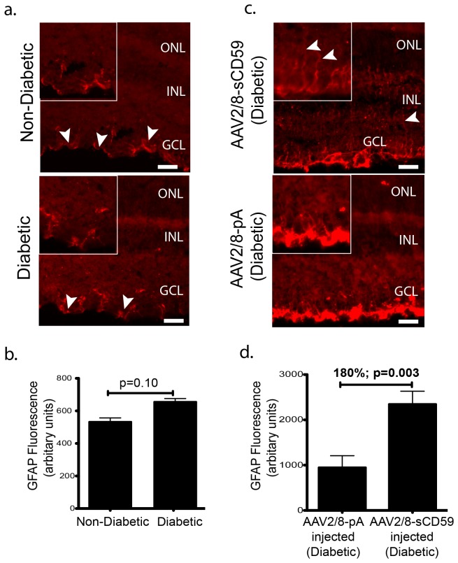
Figure 4. sCD59 Increases Retinal Müller Cell Activation in Diabetic Mice.
(a). Representative retinal sections stained for GFAP from un-injected diabetic eyes (n=7) and un-injected non-diabetic eyes (n=6) showing similar staining of astrocytes (white arrowheads) in both groups. Scale bars = 29μm. Insets: Higher magnification of stained astrocytes. (b). Quantification of GFAP staining in un-injected diabetic eyes (n=7) and un-injected non-diabetic eyes (n=6) showing no difference in the staining of the astrocytes between the two groups. (c). Representative retinal sections stained for GFAP showing increased staining of astrocytes and Müller cells in AAV2/8-sCD59 injected diabetic eyes (n=7), when compared to AAV2/8-pA injected diabetic eyes (n=6). White arrowheads indicate Müller cell processes. Scale bar = 29μm. Insets: Higher magnification. (d). Quantification of GFAP staining showing a 180% increase in staining of the glial cells in AAV2/8-sCD59 injected diabetic eyes (n=7) when compared to AAV2/8-pA injected diabetic control eyes (n=6) [p=0.006]. n represents the number of eyes. Note: GFAP staining was assessed and quantified from both central as well as peripheral retinal sections in all groups.

