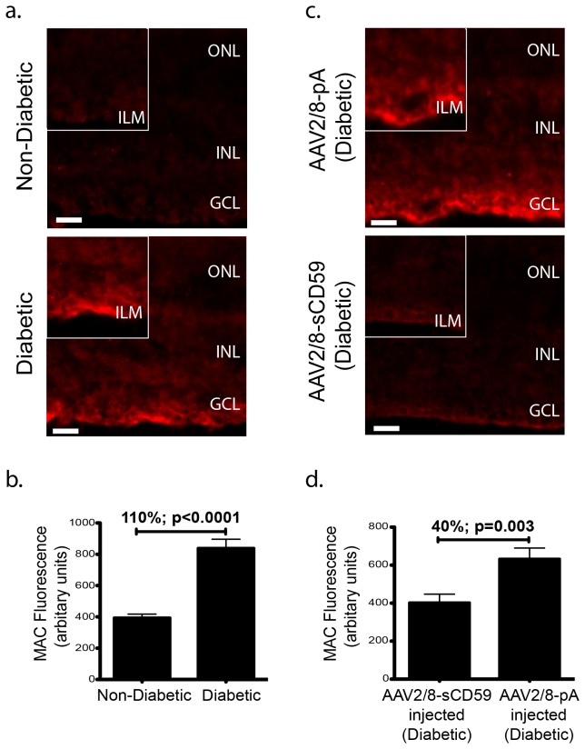
Figure 5. sCD59 Attenuates formation of Membrane Attack Complex on Retina in Diabetic Mice.
(a). Representative retinal sections stained for MAC deposition from un-injected diabetic eyes (n=7) and un-injected non-diabetic eyes (n=6) showing positive MAC staining in the inner limiting membrane in the un-injected diabetic eyes. None of the un-injected non-diabetic eyes (n=0/6) showed staining for MAC, while 43% (n=3/7) of the retinal sections from the diabetic retinas stained positive for MAC. Scale bars = 29μm. Insets: Higher magnification showing increased MAC deposition typically in the inner limiting membrane in un-injected diabetic eyes. (b). Quantification of MAC fluorescence intensity in the inner limiting membrane from un-injected diabetic eyes staining positive for MAC (n=3) showing a 110% increase in MAC deposition, when compared to un-injected non-diabetic eyes (n=6) [p<0.0001]. (c). Representative retinal sections showing reduction in MAC staining in the inner limiting membrane in AAV2/8-sCD59 injected diabetic eyes (n=7) when compared to AAV2/8-pA injected diabetic eyes (n=6). Scale bars = 29μm. Insets: Higher magnification. The reduction in MAC deposition in the AAV2/8-sCD59 injected diabetic eyes was observed as a reduced intensity of MAC staining when compared to AAV2/8-pA injected diabetic eyes. (d). Quantification of MAC fluorescence intensity in the inner limiting membrane of the retina showing a 40% reduction in MAC deposition in AAV2/8-sCD59 injected diabetic eyes (n=7) when compared to AAV2/8-pA injected diabetic eyes (n=6) [p=0.003]. n represents the number of eyes. MAC, membrane attack complex. Note: MAC staining was assessed and quantified from both central as well as peripheral retinal sections in all groups.

