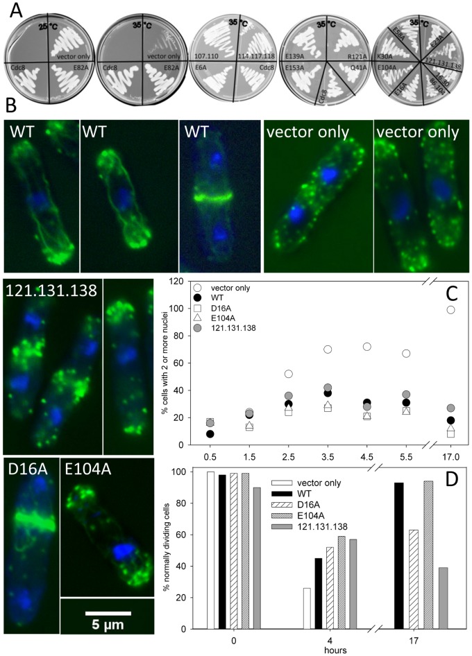
Figure 3. Proteomic screen: Rescue of cdc8-27 by pREP41X-cdc8.
A. Cdc8-27 cells were transfected with pREP41X (vector alone) or expressing wildtype or mutant Cdc8p and grown on EMM agar, lacking thiamine. The plate on the left was grown at 25°C, the four others were grown at the restrictive temperature, 35°C. B, C, D. Analysis of cells over-expressing wildtype Cdc8p and three Cdc8p mutants: D16A, E104A, and R121A.D131A, E138A in cdc8-27. Cells were grown to mid-log phase in EMM medium with thiamine, and transferred to EMM medium without thiamine and grown overnight at 25°C. Cells from the overnight culture were transferred to the restrictive temperature (35°C). Samples were taken at times after transfer and fixed. Cultures were maintained in the mid-log phase of growth. B. The cells for Alexa Fluor-488/DAPI- stained images were fixed at 4 hr (vector only, wildtype and E104A) or 27 hours (D16A, R121A.D131A.E138A) after transfer to 35°C. The images are of cells selected to show the types of observed abnormalities. C, D. The cells were fixed and stained with Calcofluor and DAPI and analyzed for the number of nuclei and the appearance of the septum in cells with two nuclei C. Percent of cells with ≥2 nuclei. At least 200 cells were counted for each time point. D. Percent normally dividing cells: Percent of binuclear septated cells showing a single, medial well-defined septum that extends from one side to the other. At least 50 cells were counted for each time point.

