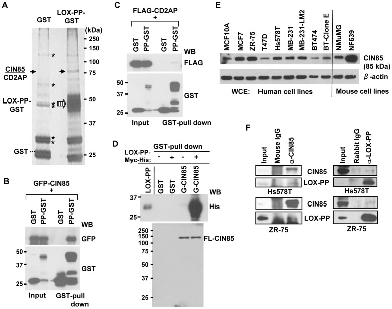
Figure 1. Identification of CIN85 and CD2AP as LOX-PP interacting proteins in breast cancer cells.
(A) Extracts from ZR-75 cells transfected with vectors expressing GST or LOX-PP-GST were precipitated with Glutathione-Sepharose 4B beads, resolved by SDS-PAGE and silver stained. The band(s) at ∼85 kDa was analyzed by LC-MS/MS mass spectrometry and identified as CIN85 and CD2AP. *, non-specific proteins. The positions of the co-precipitated CD2AP/CIN85 and LOX-PP proteins are indicated by the solid and large hatched arrows, respectively, and of GST by the dashed line. (B–C) GST or LOX-PP-GST (PP-GST) was co-expressed with GFP-CIN85 WT (B) or FLAG-CD2AP (C) in HEK293T cells, and LOX-PP associated proteins isolated by GST-pull down assays and subjected to WB for GFP (B) or FLAG (C) and GST. Input, 4% of lysates (4%). (D) Recombinant LOX-PP-myc-His (0.5 µM) was subjected to a GST-pull down assay using 0.5 µM of either GST or GST (G)-CIN85, and WB for the His or CIN85 (Calbiochem) antibody. Input, 5%. (E) Samples of whole cell extracts (10 µg) of the indicated human and mouse cells were subjected to WB for CIN85 (Upstate). (F) (Left) TX-100 extracts of Hs578T (Upper) or ZR-75 (Lower) cells were immunoprecipitated with mouse IgG or CIN85 (Upstate) antibody, and analyzed for CIN85 (Upstate) and LOX-PP. (Right) TX-100 extracts of Hs578T (upper) or ZR-75 (lower) cells were immunoprecipitated with rabbit IgG or LOX-PP antibodies, and subjected to WB.

