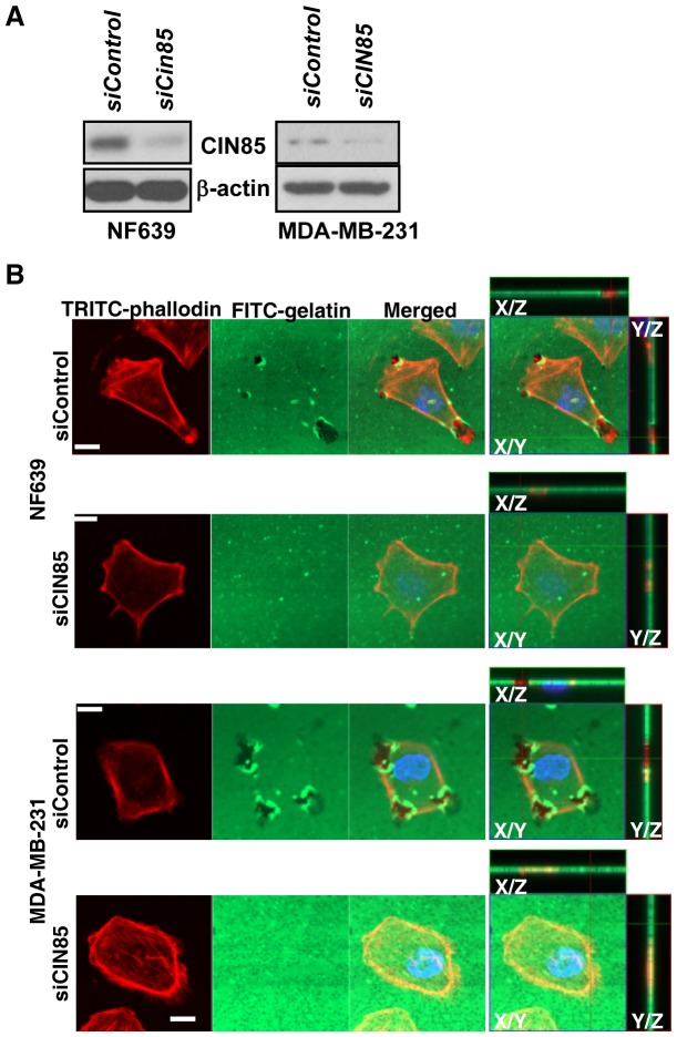
Figure 6. CIN85 promotes degradation of the extracellular matrix.
NF639 and MDA-MB-231 breast cancer cells were transfected with 20 nM of either siCIN85 or scrambled negative control siRNA (siControl). (A) After 48 h, samples of whole cell extracts (10 µg) were subjected to WB for CIN85 (Upstate: clone84) and β-actin. (B) Alternatively, cells were plated on coverslips coated with FITC-conjugated gelatin and incubated for 4 h. Cells were fixed 3.7% paraformaldehyde. F-actin and nuclei were labeled with TRITC-phalloidin (red) and Hoechst 33342 (blue), respectively. Lines indicate region of XY image projected to generate orthogonal planes XZ and YZ. Bars: 10 µm.

