
Figure 6. Protein patterns of H. polygyrus L4 larvae and H. polygyrus antigenic proteins recognized by IgG1 immune sera of BALB/c mice infected with H. polygyrus.
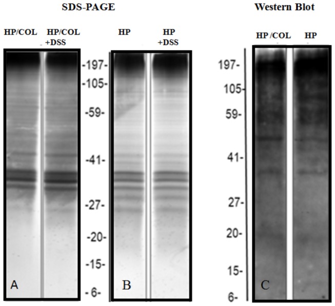
Protein patterns of L4 nematodes isolated from mice with colitis (HP/COL, A) and from control infection (HP, B) cultured in medium alone and in medium containing 5% DSS (HP+DSS; HP/COL+DSS). L4 antigen was separated by SDS-PAGE in a 4-12% gradient for 40 min at constant 200 V. Gels were silver stained. C: The blot was probed with mouse serum (1:100), followed by horseradish peroxidase-conjugated anti-mouse IgG (1:20000). The representative gel and Western blot immunedetection is shown.