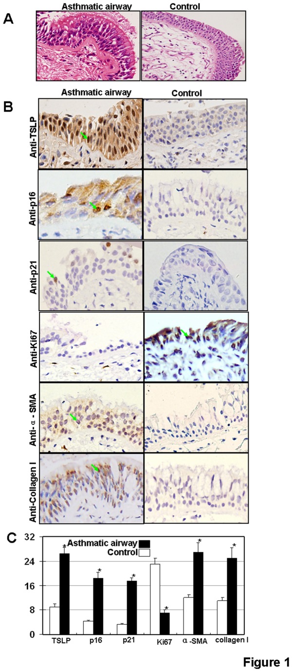
Figure 1. Protein expressions of p16 and p21 in human asthmatic airway epithelium tissue.
(A) Micrographs of histological sections of the asthmatic human airway showing the loss of epithelial integrity and wall thickening by H and E staining. Magnification 400X. (B) TSLP, p16, p21, Ki67, α-SMA and collagen I expression. Arrows indicate areas of positive expression. Magnification, 400×. (C) Bimodal H score distribution of TSLP, p16, p21, Ki67, α-SMA and collagen I immunoperoxidase reactions.

