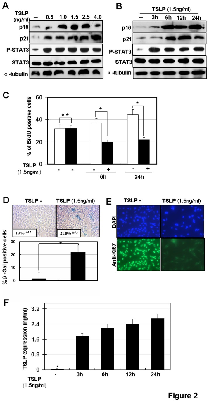
Figure 2. Cellular senescence is induced by TSLP stimulation in vitro.
(A) TSLP-induced p16 and p21 upregulation occurs in a TSLP dose-dependent manner in human bronchial epithelial BEAS-2B cells. BEAS-2B cells were stimulated with different doses of TSLP as indicated for 6h. Protein expressions of p16, p21 and phospho-Stat3 (Try705) were detected by western blotting. (B) BEAS-2B cells were stimulated by 1.5ng/ml TSLP and protein expressions of p16 and p21 were detected by western blotting. (C) BEAS-2B cells were stimulated with 1.5ng/ml TSLP then stained for BrdU. (*p< 0.05). BEAS-2B cells were stimulated with 1.5ng/ml TSLP then stained for SA-β-gal activity at 6 and 24 hours post stimulation. (D) upper panel: SA-β-gal staining; lower panel: quantification of SA-β-gal positive cells. (*p < 0.05); (E) Ki67 staining. (F) Levels of TSLP in culture media were examined by ELISA.

