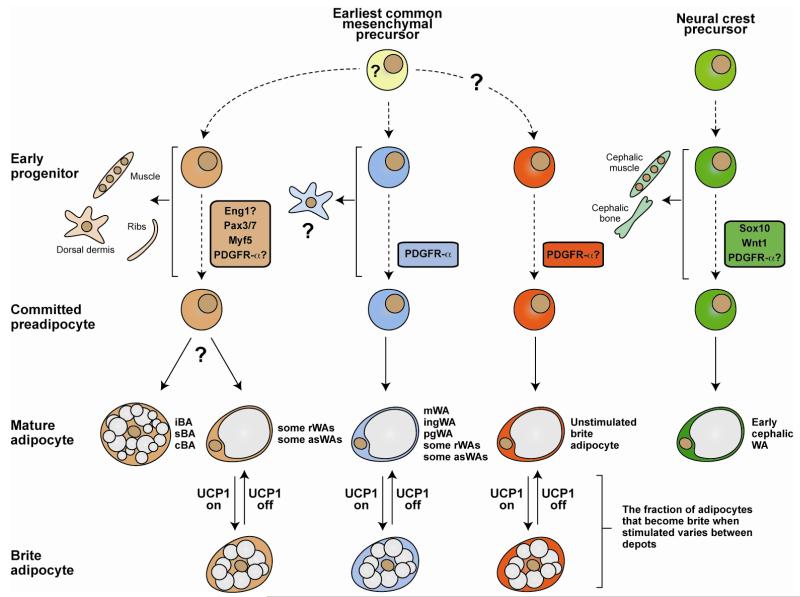
Figure 4. A model based on recent lineage tracing studies that explains some of the complexity of adipocyte ancestry.
A trending idea is that morphologically brown (multilocular), white (unilocular), and brite (interconvertible) adipocytes develop along unique lineages. However, recent studies in mice using Cre-Lox technology to irreversibly label cellular lineages argues that it is likely not this simple. An alternative view is that morphologically white adipocytes arise from multiple distinct lineages. One of these lineages is also marked by ancestral Myf5 expression and gives rise to morphologically brown adipocytes, a subset of morphologically white adipocytes (e.g. in the asWAT and rWAT), skeletal muscle cells, dermis, and ribs. Some of the Myf5-positive lineage derived white adipocytes become brite adipocytes when stimulated (e.g. Myf5-positive lineage white adipocytes in rWAT and asWAT). Other markers such as Engrailed-1 might also express early in this lineage. Whether the Myf5 lineage derived brown and white adipocytes arise from the same precursor is still unknown. Most white adipocytes in ingWAT and pgWAT develop from a Myf5-negative lineage (or possibly multiple lineages). Some of these Myf5-negative lineage white adipocytes can also become brite when stimulated (e.g. the Myf5-negative lineage white adipocytes that undergo interconversion in the ingWAT). Some brite adipocytes might also originate from a lineage distinct from the other white adipocytes although a Cre driver unique to a brite adipocyte lineage has not been found. Although white/brite adipocyte lineages are just beginning to be defined, PDGFRα appears to express in the ingWAT, pgWAT, rWAT, and mesenteric WAT lineages. Importantly, when each lineage mark actually expresses, when the lineages diverge during development, and the earliest common mesenchymal progenitor cell remain unclear and this is indicated by dotted lines and question marks. Another distinct white adipocyte lineage arises from Sox10 and Wnt1-expressing neural crest precursors in the cephalic region of young mice (notably, an unidentified lineage begins replacing neural crest derived cephalic adipocytes with age). These adipocytes label positive for PDGFR-α+ by antibody staining and therefore likely would trace with PDGFR-Cre, although this has not been shown. In sum, the emerging lineage tracing data presents a complex picture of adipocyte origins. iBA, sBA, cBA = interscapular, subscapular, and cervical brown adipocyte; asWA, rWA, pgWA, ingWA = anterior subcutaneous, retroperitoneal, perigonadal, inguinal white adipocyte. For more detailed information see [50, 51, 83, 100, 103, 114, 116, 127].

