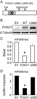
Figure 5.
DNA binding domain of FOXO1 is required to suppress basal and GnRH-induced Fshb gene expression. (A) Diagram illustrating FOXO1-ΔDBD (Δ208–220). (B) LβT2 cells were transfected with pcDNA3 empty vector (EV), pcDNA3-FOXO1 (WT), or pcDNA3-FOXO1-ΔDBD for 6 hours, then switched to serum-free media. Twenty-four hours after transfection, the cells were harvested. Western blot analysis was performed on whole cell extracts using FOXO1 and β-Tubulin primary antibodies and a horseradish peroxidase–linked secondary antibody. A representative image is shown. (C–D) The −1000 murine Fshb-luc reporter was transfected into LβT2 cells along with EV, FOXO1, or FOXO1-ΔDBD, as indicated. After overnight incubation in serum-free media, cells were treated for 6 hours with 0.1% BSA or 10 nM GnRH. The results represent the mean ± SEM of three experiments performed in triplicate and are presented as basal transcription relative to empty vector (C) or fold GnRH induction relative to the vehicle control (D). *, Fshb-luc transcription is significantly repressed by FOXO1 compared to EV or FOXO1-ΔDBD using one-way ANOVA followed by Tukey's HSD post-hoc test.

