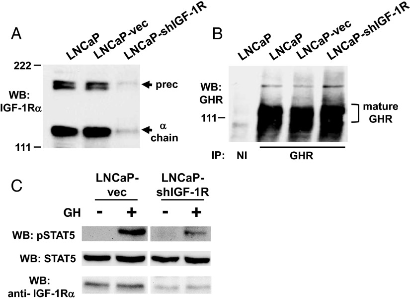
Figure 3.
shRNA-mediated silencing of IGF-1R in LNCaP cells results in reduced GH-induced STAT5 phosphorylation. A–C, Characterization of pools of LNCaP human prostate cancer cells stably transfected with either vector only (LNCaP-vec) or a vector driving expression of an shRNA directed at IGF-1R (LNCaP-shIGF-1R). A, Equal amounts of protein from detergent cell extracts of parental LNCaP, LNCaP-vec, and LNCaP-shIGF-1R cells were resolved by SDS-PAGE and immunoblotted with anti-IGF-1Rα antibody. Arrows indicate the positions of the IGF-1R precursor and α-chain. Note the similarity of abundance of IGF-1R in LNCaP and LNCaP-vec cells but marked reduction in IGF-1R in LNCaP-shIGF-1R cells. WB, Western blotting. B, Equal amounts of protein from detergent cell extracts of parental LNCaP, LNCaP-vec, and LNCaP-shIGF-1R cells were immunoprecipitated with anti-GHR cytoplasmic domain antiserum or nonimmune (NI) serum. Eluates were resolved by SDS-PAGE and immunoblotted with anti-GHR. Bracket indicates the position of the mature GHR. Note similarity of GHR abundance among the three cell lines. C, Serum-starved LNCaP-vec and LNCaP-shIGF-1R cells were treated with GH (+; 250 ng/ml) or vehicle (−) for 15 minutes. Detergent cell extracts were resolved by SDS-PAGE and serially immunoblotted with anti-pSTAT5, anti-STAT5, and anti-IGF-1Rα. Note marked reduction in GH-stimulated STAT5 phosphorylation in LNCaP-shIGF-1R cells compared with LNCaP-vec cells.

