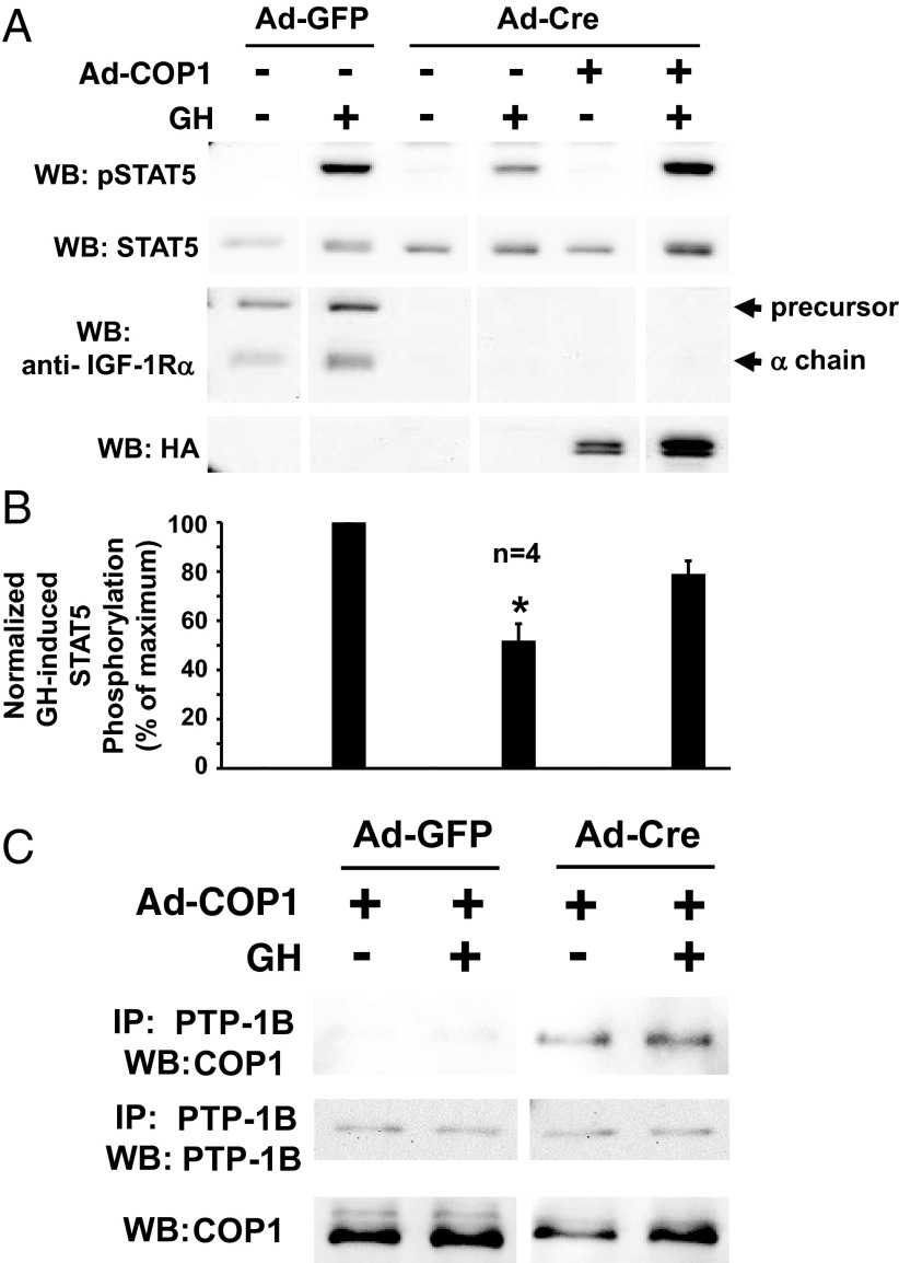
Figure 6.
COP1 rescues the diminished GH-induced STAT5 phosphorylation resulting from IGF-1R depletion and COP1-PTP-1B interaction is enhanced in IGF-1R-depleted osteoblasts. A and B, Primary osteoblasts were infected with Ad-Cre vs Ad-GFP, as indicated, and Ad-COP1 (+) or Ad-GFP (−), as indicated. Serum-starved cells were treated with GH (+; 250 ng/mL) or vehicle (−) for 10 minutes. Detergent cell extracts were resolved by SDS-PAGE and serially immunoblotted with anti-pSTAT5, anti-STAT5, anti-IGF1Rα, and anti-HA (to detect HA tagged COP1 proteins). A, Representative immunoblots. WB, Western blotting. B, Densitometric quantitation of pSTAT5/STAT5 signals from GH-treated samples from four independent experiments (including that shown in A). In each experiment, the maximum signal was considered 100%. Data are plotted as mean ± SE. *, P < .02 for comparison of Ad-Cre-infected, non-Ad-COP1-infected, GH-treated group with either the Ad-GFP-infected, non-Ad-COP1-infected, GH-treated group or the Ad-Cre-infected, Ad-COP1-infected, GH-treated group. C, Coimmunoprecipitation of COP1 with PTP-1B. Primary osteoblasts were infected with Ad-Cre vs Ad-GFP, as indicated, and Ad-COP1 (+), as indicated. Serum-starved cells were treated with GH (+; 250 ng/mL) or vehicle (−) for 10 minutes. Detergent cell extracts were either immunoprecipitated with anti-PTP-1B or not immunoprecipitated. Eluates or extracts were resolved by SDS-PAGE and immunoblotted with anti-COP1 or anti-PTP-1B, as indicated. IP, immunoprecipitation; WB, Western blotting.

