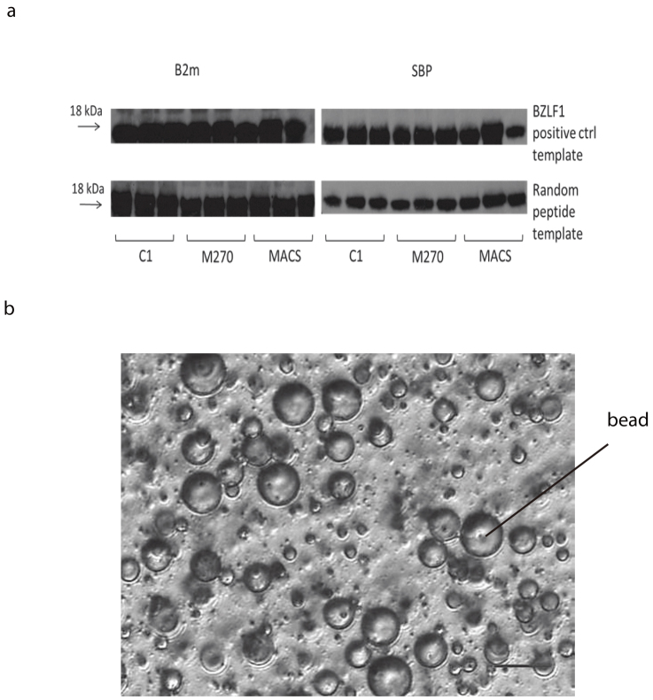
Figure 3.
(a) emIVTT on different types of beads. Cropped western blot results showing protein-DNA-beads attaching with either EBV BZLF1 positive control template (known peptide sequence) on upper row, or templates containing randomised peptide sequences (lower row). Full gel is available as supplementary information. The left column shows the results of human β-2-microglobulin (β2m) staining and the right shows SBP staining. Each type of beads was labelled with three different concentrations of biotin labelled primers attaching on the beads, e.g. for C1 column, 1000 pmol, 500 pmol, 250 pmol primers were attached on 1 mg of beads (6–7 × 10⁷). Figure 3(b) The 40× inverted microscopic image of a water-in-oil emulsion mixture with Dynabead C1. A successful water-in-oil emulsion has diameter of 10-15 μm and contains a single magnetic bead in the aqueous phase. The scale bar represents 20 μm.

