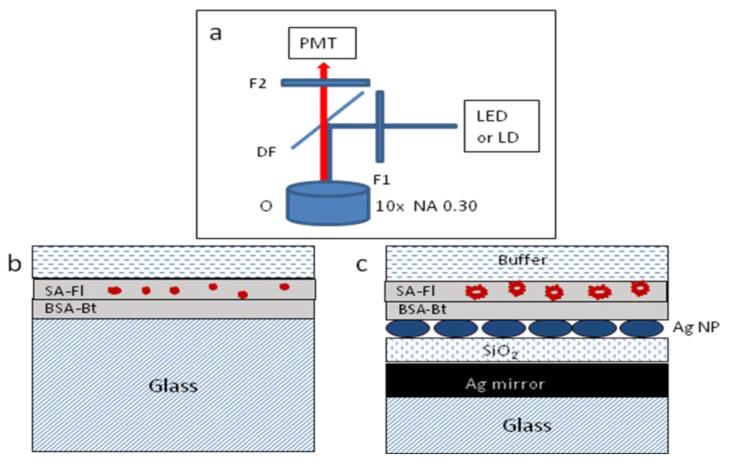
Figure 1.
Schemes of epifluorescence (a), geometrical configuration of reference sample (b), and multilayered substrate (c) with an immobilized layer of streptavidin-fluorophore (SA-Fl) on the layer of BSA-Bt. The specifics about the metal and dielectric layer thicknesses are within the text. The thicknesses in the figure are not to scale. The abbreviations of F1, F2, DF, and O are for the excitation, emission, dichroic filters, and microscope objective, respectively.

