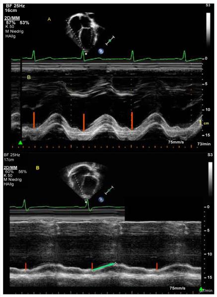
Figure 7.
Apical 4-chamber view. (A) The white broken line indicate M-Mode cursor placement at the tricuspid lateral annulus. Representative M-Mode image of the tricuspid annular plane systolic excursion (TAPSE) in a patient with normal right and left ventricular function. The absolute longitudinal displacement measure is shown as the red line. The yellow arrow marks the upper and lower measure point of one centimeter (cm). (B) Representative M-Mode image of the tricuspid annular plane systolic excursion (TAPSE) in a 17 year old patient with TOF and a decreased TAPSE. The absolute longitudinal displacement measure is shown as the red line. The green arrow shows the decreased TAPSE value and flat course of the excursion.

