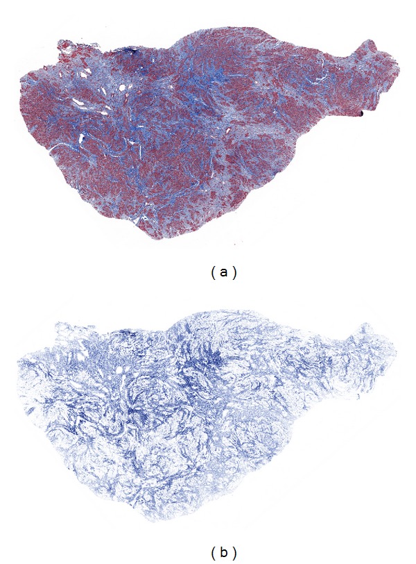
Figure 1.

Image analysis of fibroid collagen content. By light microscopic examination of the H&E-stained section of this tumor, the collagen content was estimated to be more than 10% and less than 50% and thus to fall into the phase 3 category. The image on the left (a) shows the Masson's trichrome-stained section of this tumor, while that on the right (b) depicts the markup image of the same section in which only the blue-stained collagen has been colocalized, allowing for quantitation of the percent of collagen, which was found to be 38.4%.
