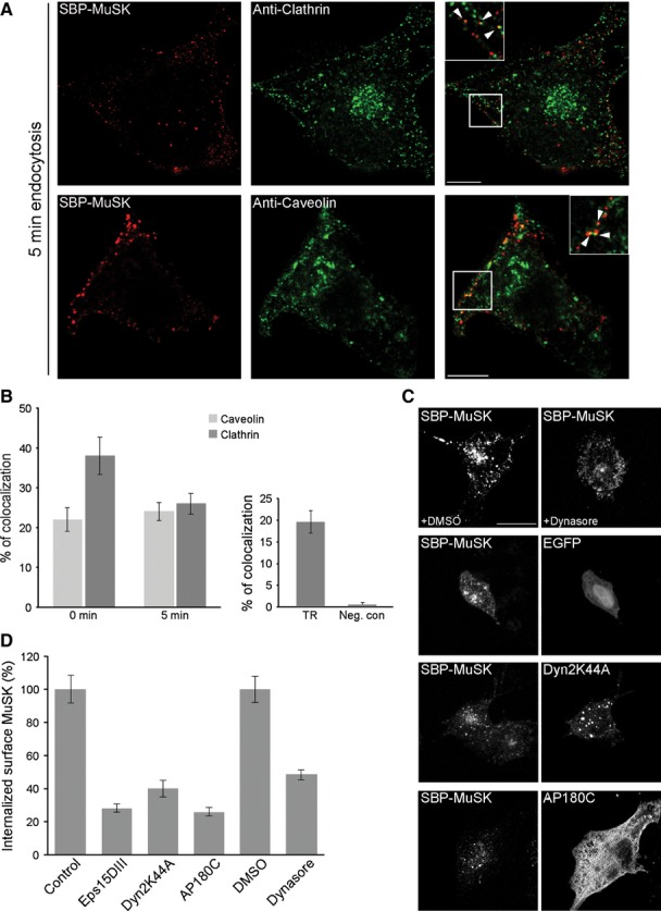
Fig 2.

MuSK internalization proceeds via a clathrin-dependent pathway. (A) To determine whether surface MuSK internalizes via clathrin-or caveolin-positive routes, COS-7 cells were transiently transfected with SBP-MuSK. Surface MuSK was stained with Cy3-conjugated streptavidin (red) at 4 °C followed by incubation at 37 °C for 5 min. Endogenous clathrin and caveolin were visualized by antibody staining. MuSK partially colocalizes with these markers (arrowheads in insets). Scale bar = 25 μm. (B) Quantification of MuSK/clathrin, MuSK/caveolin and MuSK/transferrin colocalization using a threshold-and object-based colocalization analysis (as described in the Materials and methods). Colocalization of MuSK with clathrin and caveolin was analyzed at 0 and 5 min of endocytosis. Transferrin and MuSK colocalization was analyzed at 5 min of endocytosis. The peroxisomal marker PTS2-GFP was used as a negative control. Error bars indicate the SEM (n ≥ 19; from at least two independent experiments). (C) To determine whether dynamin or clathrin are involved in MuSK internalization, COS-7 cells were either treated with the dynamin specific blocker dynasore or co-transfected with GFP-tagged Dyn2K44A or Myc-tagged AP180C. Surface MuSK was stained with streptavidin conjugated-DyLight 649 (red) at 4 °C followed by incubation at 37 °C for 30 min and subsequent stripping of the remaining surface MuSK molecules. Scale bar = 25 μm. (D) To quantify the blockage of MuSK internalization, COS-7 cells were transiently transfected with SBP-MuSK alone or, as indicated, together with AP180C, Eps15DIII or Dyn2K44A. Cells treated with dimethylsulfoxide (DMSO) or dynasore, were stained with streptavidin-conjugated DyLight 649 followed by incubation at 37 °C for 5 min. The remaining surface staining was stripped off and cells were harvested for intracellular fluorescence detection by FACS. A quantification of internalized MuSK is shown. The control sample denotes GFP-positive cells presenting a streptavidin signal. The dimethylsulfoxide sample represents streptavidin-positive cells treated with the solvent dimethylsulfoxide only. Error bars indicate the SEM (n ≥ 5).
