Fig 3.

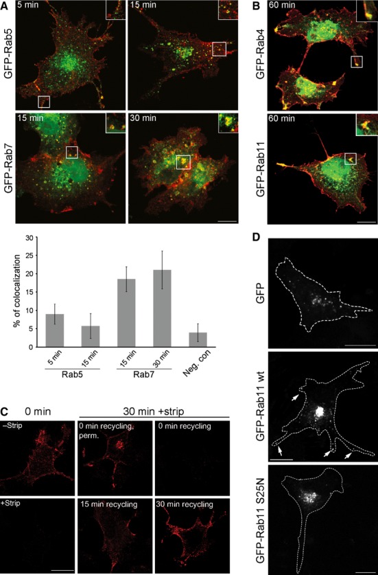
MuSK is transported in Rab7-and Rab4-/11-positive endosomes. (A) To determine whether classical endosomal markers are involved in MuSK trafficking, COS-7 cells were co-transfected with SBP-MuSK together with either GFP-tagged Rab5 (early endosomal compartments, EEA1 positive) or with GFP-tagged Rab7 (late endosomal pathway). Surface MuSK were labelled with DyLight 649-conjugated streptavidin (red) at 4 °C followed by incubation at 37 °C for different time periods. Lower panel: quantification of the colocalization of MuSK with Rab5 or Rab7 after different time points using a threshold-and object-based colocalization analysis (as described in the Materials and methods). MuSK/Arf1 was used as a negative control (neg. con). Error bars indicate the SEM (n ≥ 8). (B) COS-7 cells were co-transfected with SBP-MuSK together with either GFP-tagged Rab4 (early recycling) or with GFP-tagged Rab11 (late recycling). Surface MuSK were labelled with DyLight 649-conjugated streptavidin (red) at 4 °C followed by incubation at 37 °C for 60 min. Magnified insets demonstrate a colocalization between MuSK and Rab4 or Rab11. (C) COS-7 cells were transiently transfected with HA-MuSK, stained with an antibody against the extracellular HA-tag followed by incubation at 37 °C for 30 min. The remaining surface antibody staining was stripped off and cells were reincubated at 37 °C for 15 and 30 min (recycling). Cells were fixed and stained with secondary antibodies. Recycled MuSK is detectable at the cell membrane after 15 min. Stripping efficiently removed bound antibodies because no surface MuSK was detectable after 0 min of recycling. Perm, permeabilized. (D) COS-7 cells were co-transfected with either pEGFP, GFP-tagged Rab11 wt or GFP-tagged Rab11 S25N. Surface MuSK was labelled with Cy3-conjugated streptavidin at 4 °C followed by incubation at 37 °C for 120 min. The expression of Rab11 wt increases the recycling of MuSK to the plasma membrane (arrows). Magnified structures are shown as insets. Scale bars = 25 μm.
