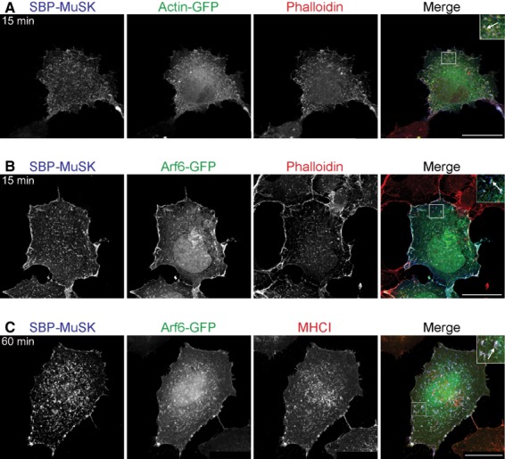
Fig 4.

MuSK localizes to structures rich in actin and Arf6. COS-7 cells were transiently transfected with SBP-MuSK. Surface MuSK was labelled with DyLight 649-conjugated streptavidin (blue) at 4 °C followed by incubation at 37 °C for 15 or 60 min. (A) MuSK internalization was visualized in COS-7 cells, which were co-transfected with GFP-tagged actin. The actin cytoskeleton was stained with rhodamine-conjugated phalloidin (red) after cell fixation. (B) MuSK internalization was visualized in COS-7 cells co-transfected with a GFP-tagged Arf6 and stained with phalloidin after cell fixation (red). (C) HeLa cells were transiently transfected with SBP-MuSK together with Arf6-GFP, followed by staining with an antibody against the endogenous Arf6 marker protein MHCI. Magnified structures demonstrating colocalization (arrows) are shown as insets. Scale bar = 25 μm.
