
Figure 6.
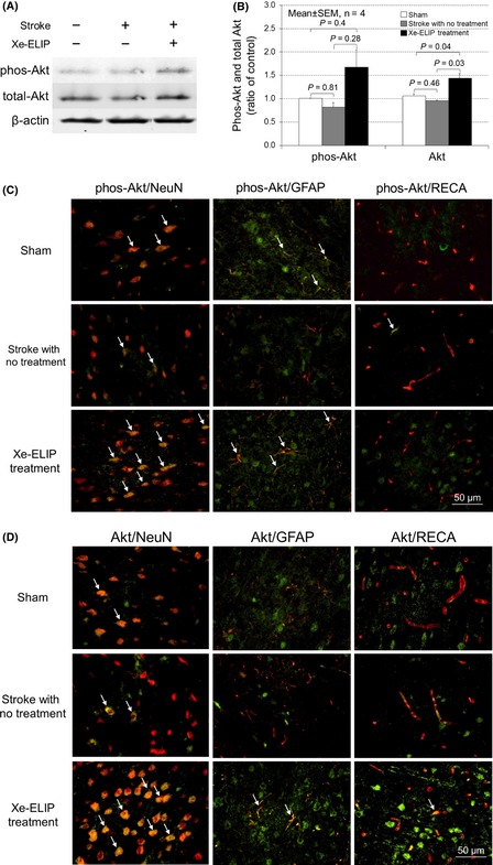
Phosphor‐Akt and total Akt expression and colocalization with neurons, astrocytes, and endothelial cells. Western blot analysis of Akt expression (A) and quantitation (B). Double immunofluorescence staining for colocalization of phosphor‐Akt (C) and total Akt (D) (green) with NeuN (red), glial fibrillary acidic protein (red), or rat endothelial cell antigens (red) in the penumbral region of the brain tissue at 24 h poststroke onset. Phospho‐Akt immunoreactivity increased in both neuronal cells and astrocytes/glia cells at 24 h after Xe‐containing echogenic liposomes treatment, but not in the endothelial cells.