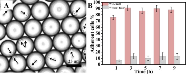
Figure 5.

(A) Representative bright-field image of Jurkat E6.1 cells (indicated by arrows) in the cRGD-functionalized nanostructured droplets 6 h after their creation. (B) Quantification (adherent cell %) of Jurkat E6.1 cell adhesion on cRGD-functionalized (pink, left bars) and nonfunctionalized (gray, right bars) nanostructured droplets. Data are presented as means ± standard errors of the mean (n = 5).
