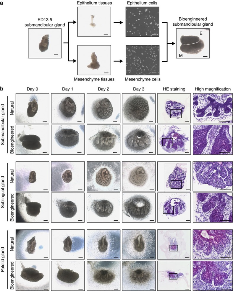
Figure 1. Generation of a bioengineered salivary gland germ.
(a) Phase-contrast images of the ED13.5 submandibular gland germ, tissues, dissociated single cells and the bioengineered submandibular gland germ, which was reconstituted using the organ germ method. E, epithelial cells; M, mesenchymal cells. Scale bar, 200 μm. (b) Phase-contrast images of natural (upper) and bioengineered (lower) salivary gland germs, including the ED13.5 submandibular gland (top columns), the ED14.5 sublingual gland (middle columns) and the ED14.5 parotid gland (bottom columns) on days 0, 1, 2 and 3 of organ culture. The salivary glands were analysed by haematoxylin and eosin staining on day 3 of organ culture. Scale bar, 200 μm.

