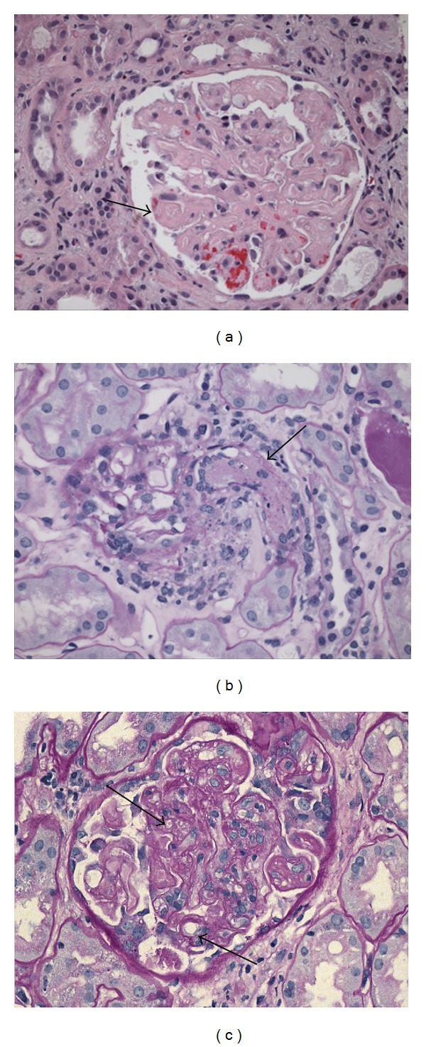
Figure 1.

Light micrographs showing TMA. (a) Patient 1: H&E 20x: glomerulus with consolidated appearance caused by swelling of endothelial cells (endotheliosis). (b) Patient 2: PAS, 20x: glomerulus with an arteriole occluded by a thrombus. (c) Patient 3: PAS, 40x: mesangiolysis and double contours.
