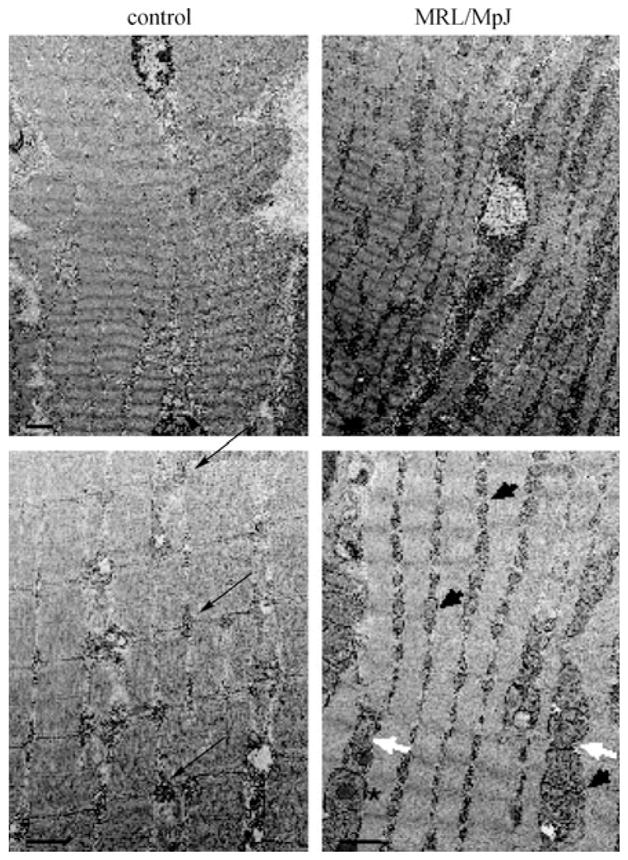
Figure 1.

Electron micrographs demonstrating aberrant mitochondria in the MRL/MpJ mouse strain quadriceps, compared to the DBA/2J control strain. Note the standard arrangement of interfibril mitochondrial pairs in the control quadriceps (black arrows) and the increased number of mitochondria at this anatomical position in the MRL/MpJ strain (black arrowheads). Some of the MRL/MpJ mitochondria also demonstrate abnormal shapes (white arrows) and lipid-like inclusion bodies (asterisk). Top panels’ bar is 2 μm, bottom panels’ bar is 1 μm.
