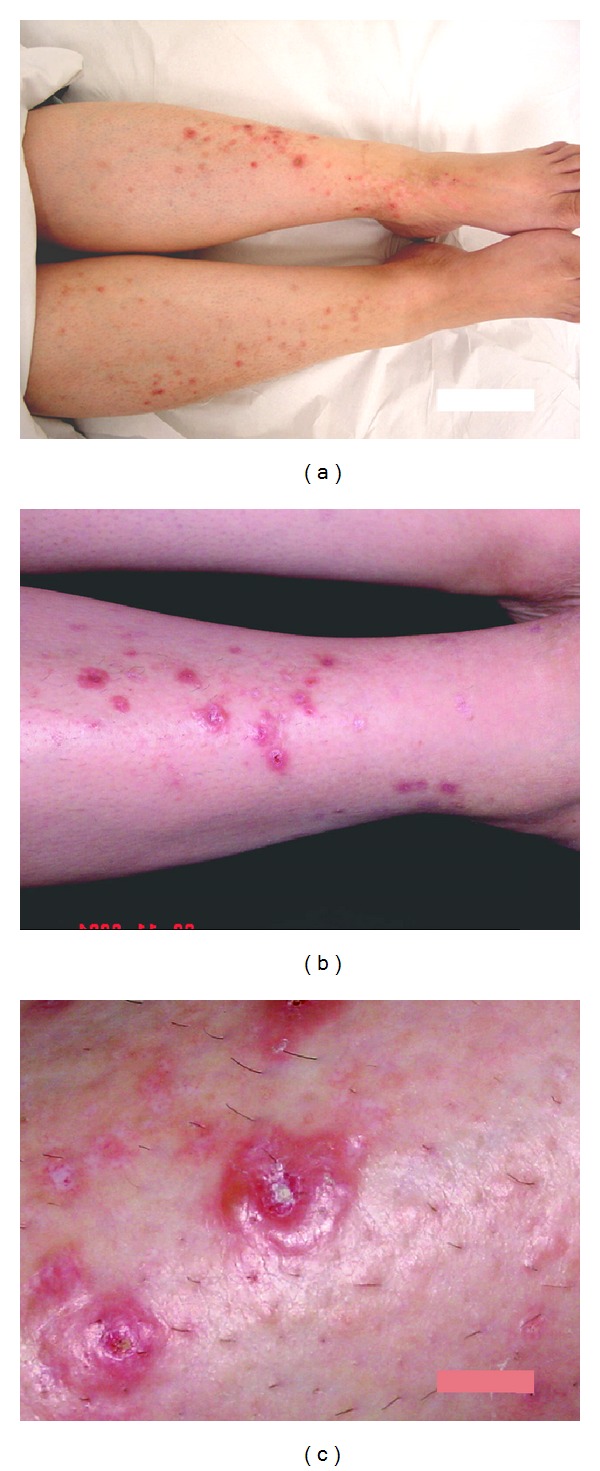
Figure 1.

(a) The distribution of the lesions, bilateral and almost symmetrical, on both lower limbs. (b) Higher magnification showing the vesicular lesions lying on erythematous base and the fine white scars in healed lesions. (c) A close-up view of the vesicular lesions showing tense blistering with central crustations, on the right side of the photo a superficial white scar of healed lesion.
