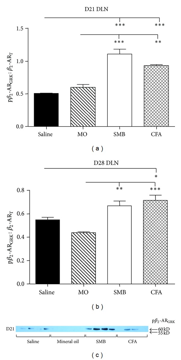
Figure 9.

Expression of the pβ 2-ARGRK/β 2-ART in DLN cells from rats challenged with CFA, SMB, or MO compared with Saline-treated controls on D21 (a) and D28 (b). No difference in pβ 2-ARGRK/β 2-ART was observed between the MO- and Saline-treatment groups at either time point. Expression of pβ 2-ARGRK/β 2-ART was increased in CFA-, SMB- compared with MO-challenged and Saline-treated rats on D21 and D28. DLN cells were harvested, lysed, and proteins resolved by SDS-PAGE. Cellular extracts were probed with an antibody against GRK phosphorylated Ser355/Ser356 of the β 2-AR. A western blot is shown (c) that is representative of the blots seen within each treatment. The data were normalized to β-actin, and pβ 2-ARGRK expression was normalized to β 2-ART. Data are expressed as a mean pβ 2-ARGRK/β 2-ART ± SEM with an n of 8 rats per treatment group. Data were analyzed using one-way ANOVA with Bonferroni posthoc testing (**P < 0.01; ***P < 0.001).
