
Figure 2.
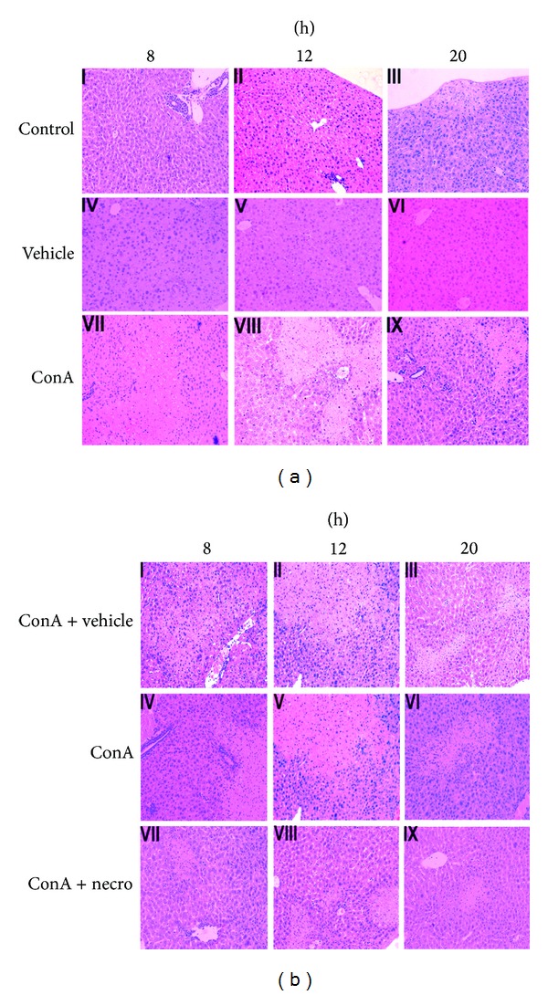
(a) Photomicrographs of representative livers collected 8, 12, and 20 h after ConA injection, stained with H&E (magnification ×200). The control animals received NSS alone ((a) I–III). The vehicle-injected group received DMSO only, without ConA ((a) IV–VI). The mice in the ConA group were injected in the tail vein with ConA for at 8, 12, or 20 h. H&E, ×200 magnification ((a) VII–IX). These experiments were repeated three times, and the same results are shown. (b) Photomicrographs of representative livers collected at 8, 12, and 20 h after ConA injection, stained with H&E (magnification ×200). The vehicle group was intraperitoneally injected with DMSO 1 h before ConA challenge ((b) I–III). ConA-injected control animals received ConA only ((b) IV–VI). The Nec-1-pretreated group was intraperitoneally injected with Nec-1 1 h before ConA challenge ((b) VII–IX). These experiments were repeated three times with the same results.