
Figure 6. Down-regulation of ERLIN2 reduces cancer cell proliferation rates.
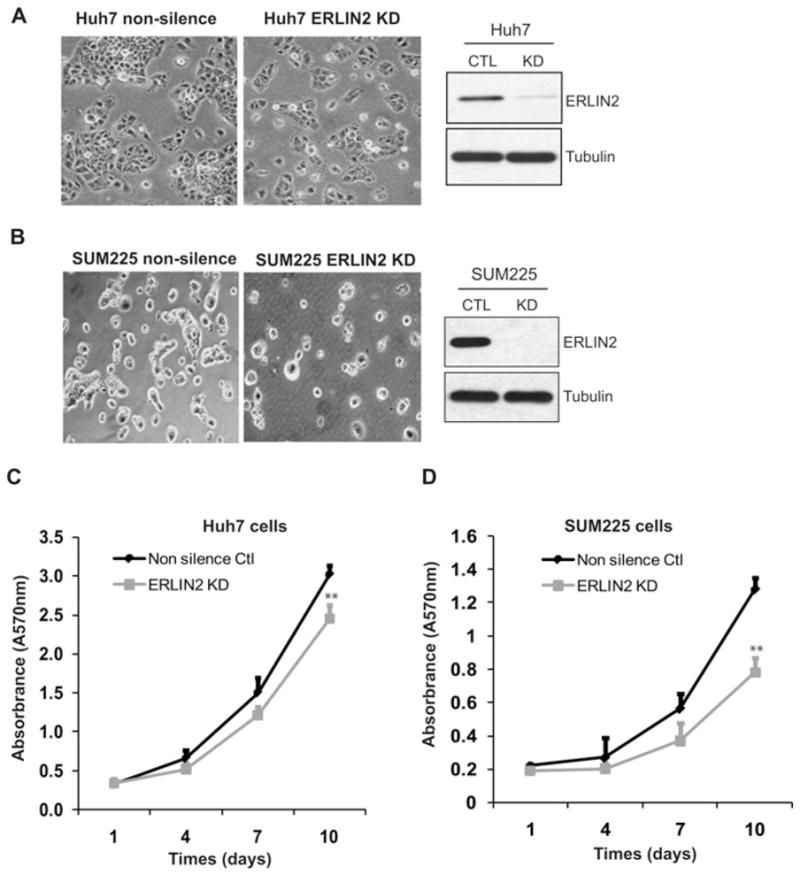
(A) Left-hand panel, morphology of ERLIN2 knockdown and control Huh-7 cells. Magnification is ×400. Right-hand panel, levels of ERLIN2 protein in knockdown (KD) and control (CTL) Huh-7 cells were determined by Western blot analysis. (B) Morphology of ERLIN2 knockdown and control SUM225 cells. Magnification is ×400. Levels of ERLIN2 protein in knockdown and control SUM225 cells were determined by Western blot analysis. (C and D) Cell proliferation assays with the stable ERLIN2 knockdown Huh-7 (C) or SUM225 (D) cells and control cells. On day 1, the same number of ERLIN2 knockdown or control cells was seeded (5000 cells per well). The cell proliferation rate at each time point was represented by a 570-nm absorbance reading determined by MTT assay. Results are means ± S.E.M. (n = 3 biological samples). **P < 0.01.