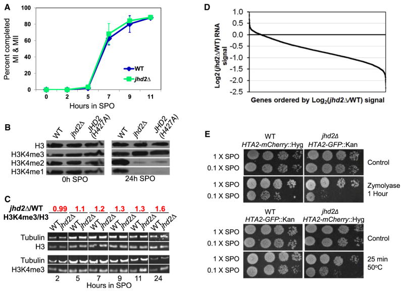
Figure 1. JHD2 Functions during Sporulation to Control Global Transcript Accumulation, H3K4 Demethylation, and Gamete Fitness.
(A) Meiotic progression of WT and jhd2Δ cultures was quantified as percent of cells that have completed meiosis I and II. Shown are averages of three independent experiments. Error bars indicate 1 SD.
(B) Western blotting was used to detect abundance of H3 and H3K4me in WT and jhd2Δ strains at the indicated time points of sporulation. Membranes were stripped and reprobed with the indicated antibodies.
(C) Quantitative western blotting was performed on aliquots from sporulating WT and jhd2Δ cultures using the indicated primary antibodies. After normalization to tubulin signal, the H3-3meK4/total-H3 ratio of jhd2Δ compared with WT was calculated and is shown. See also Figure S1.
(D) Affymetrix tiling arrays were used to measure RNA abundance from WT and jhd2Δ spores (20 hr) at a resolution of 4 bp. Normalized intensities for protein coding genes were plotted for jhd2Δ cells relative to WT and ordered by decreasing fold change for all annotated genes. For comparison, see Figure S1A and Table S1 for relative WT and jhd2Δ RNA abundance data in vegetative cells.
(E) WT and jhd2Δ mutants of the indicated genotypes were cosporulated in 1× or 0.1× SPO medium. Sporulated cultures were treated with zymolyase or 55°C heat and were assayed for survival by monitoring postgermination growth.

