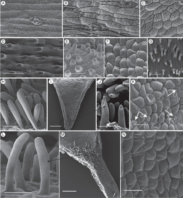
Fig. 3.
Scanning electron micrographs of the spur epidermis of selected nectarless Disa species. (A) Oblong epidermis cells with longitudinal striations and a malformed stoma near the spur tip of D. harveiana subsp. longicalcarata. (B) Mature and immature stoma near the spur tip of D. tysonii. (C) Thick, irregular cuticular striations characterize the entire spur epidermis of D. stachyoides. (D) Thickly cuticularized, papillate cells line the entire spur of D. nervosa. (E) Papillate epidermis cells line the entire spur of D. obtusa subsp. hottentotica. (F, G) Cells lining the spur (F) and papillae (G) in spur entrance of D. obliqua subsp. obliqua. (H) Club-shaped unicellular trichomes of D. cephalotes subsp. cephalotes. (I) Spur overview of D. caulescens. (J) Close-up of papillae in spur tip. The tips of the frontal two papillae were damaged during spur sectioning. (K) Scattered stomata in D. racemosa (indicated by arrows). (L–N) D. uncinata. (L) Spur epidermis; (M) overview of spur section showing field of trichomes in spur entrance; (N): close-up of unicellular trichomes. Scale bars: (A–F, H, J, L, N) = 50 μm; (G, K) = 100 μm; (I, M) = 0·5 mm.

