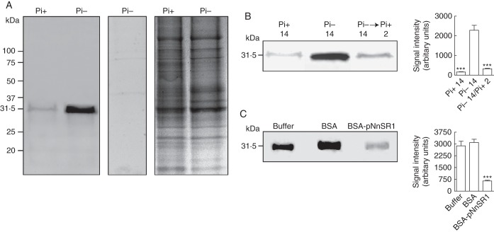
Fig. 3.
Expression of class III NnSR1 protein induced in Nicotiana alata roots under Pi deprivation. NnSR1 induction was assayed by Western blot in roots subjected to Pi deprivation for 14 d. Anti KLH-pNnSR1 serum was used as probe. (A) Induction of a 31-kDa protein in root homogenates of Pi-starved plants. Controls using preimmune serum (central panel) and Coomassie blue staining (left panel) are shown. The result is representative of three independent experiments. Similar result was obtained using the anti-BSA-pNnSR1 serum. (B) Root homogenates assayed after Pi-starved plants were transferred to Pi-supplied medium for 2 d. (C) Root homogenates assayed after preincubation of anti KLH-pNnSR1 serum with buffer and with 3 × 10⁻¹⁰ mol of BSA and 3 × 10⁻¹⁰ mol of BSA-conjugated pNnSR1. Thirty micrograms of protein was loaded in each lane. Signal intensity values represent the mean ± s.e.m. of three independent experiments. The data in (B) and (C) were statistically analysed using one-way ANOVA followed by Tukey test: ***P < 0·001. Pi-, Pi-deficient medium; Pi+, Pi-sufficient medium.

