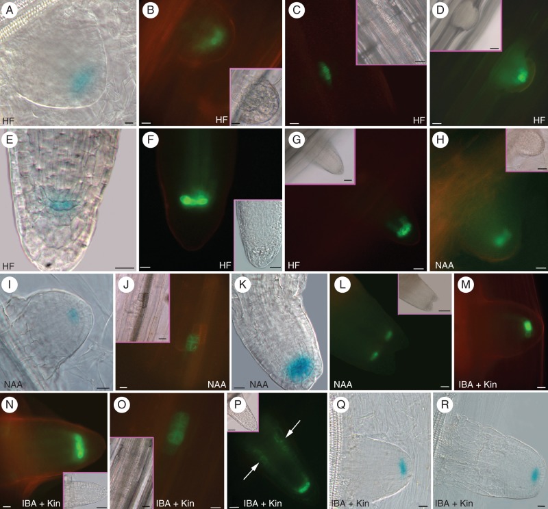
Fig. 2.
Expression of PR/LR QC markers during AR formation in Col seedlings grown for 14 d under various hormonal treatments. (A–G; HF) QC25::GUS (A), pAGL42::GFP (B) and pWOX5::GFP (D) in the QC at stage VII, and pWOX5::GFP at stage II (C). QC25::GUS (E), pAGL42::GFP (F) and pWOX5::GFP (G) in the QC, and lateral initials (G) of emerged ARs. (H–L; NAA) pAGL42::GFP (H) and QC25::GUS (I) in the QC of stage VII ARPs. pWOX5::GFP at stage II (J), QC25::GUS at the tip of a regular AR (K) and pAGL42::GFP in the twin tip of a fasciated AR (L). (M–R; IBA + Kin) pAGL42::GFP in the QC of not yet emerged (M) and emerged (N) ARPs. pWOX5::GFP at stage II (O), and in the QC, lateral initials and pericycle cells forming LRs (arrows) in emerged ARPs (P). QC25::GUS in the QC at stage VII (Q), and in emerged ARPs (R). Insets in fluorescence pictures show corresponding bright-field images. Scale bars: = (A, B and inset, E, F, H–K, M–O, R) 10 µm; (C, D, G, L, P–Q, and insets in C, F, J, N–O) = 20 µm; (insets in D, G, H) = 30 µm; (inset in P) = 40 µm; (inset in L) = 100 µm.

