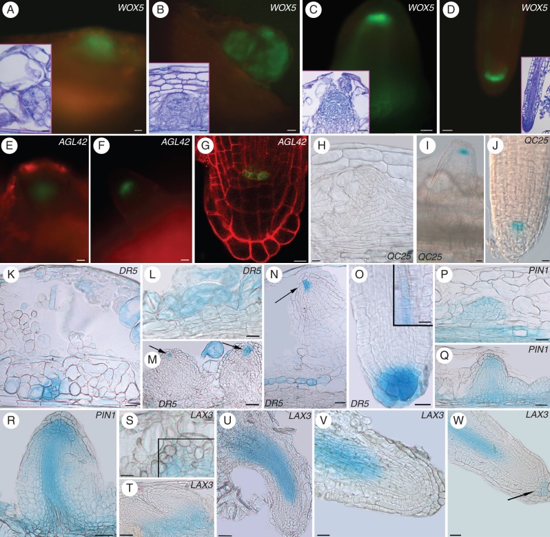
Fig. 6.
Expression of QC markers (A–J), auxin monitoring (K–O), and auxin efflux (P–R) and influx (S–W) gene expression during AR formation in IBA + Kin-cultured TCLs. (A, B) WOX5 expressed in meristematic cell clusters (A) and root meristemoids (B). Corresponding light microscopy images are shown in the insets. (C, D) Emerging ARPs (C) and apices of elongated ARs (D) showing WOX5 in the QC and lateral initials. Corresponding light microscopy images are shown in the insets. (E) Early-domed ARPs showing the appearance of pAGL42. (F) Emerging ARPs with pAGL42 expression in the QC. (G) Confocal microscopy image of mature AR apices with pAGL42::GFP signal in the QC. (H) No expression in early-forming ARPs of QC25::GUS TCLs, but expression in the QC of emerged ARPs (I) and elongated ARs (J). (K) Meristematic cell clusters with DR5::GUS expression. (L) Detail of a meristemoid showing signal. (M, N) Not yet protruded ARPs showing DR5 expression in the tip (arrows in N). (O) DR5::GUS expression in the niche and cap of mature ARs. The inset shows expression in the forming vasculature. (P) PIN1 expression in early-domed ARPs, corresponding to stage VII in planta, and endodermis-derived cells at the base. (Q) Not yet emerged ARPs showing PIN1 signal, mainly in the tip and forming vasculature. (R) Protruded ARP with PIN1 expression in the vasculature, procambium and apex. (S) LAX3 expression in forming meristemoids (square). (T) Not yet protruded ARPs with LAX3 expression at the base. (U, V) Elongating ARPs after protrusion, with LAX3 expression in the vasculature, but not in the apex. (W) Mature AR with LAX3 signal in the developing vasculature and some cap cells (arrow). Insets in A–D, toluidine blue section staining. Scale bars: (insets in A and C, G) = 10 µm; (A, C–F, H, J, O, inset in O, V, W) = 20 µm; (inset in B, I, T) = 30 µm; (inset in D, K–M, S, U) = 40 µm; (N, P–R) = 50 µm; (B) = 100 µm.

